Medicilon in one place
Dive Deeper
Corporate News
- Corporate News
Medicilon and Hailu Biotech of Yangtze River Pharmaceutical Group Forge Strategic Cooperation to Accelerate New Drug R&D and Global Outreach
- Corporate News
Medicilion Congratulates MediLink Therapeutics on Licensing B7H3 ADC to Roche
- Corporate News
Building a New-Drug Expressway Together, Medicilon 2025 Year-End Review
- Corporate News
Medicilon Congratulates AI-Driven Biotech Insilico Medicine on Its HKEX Debut in Hong Kong’s Largest Biotech IPO of 2025
- Corporate News
Efficient Collaboration Earns High Praise: RayThera Honors Medicilon with “Excellent Partnership Award”
- Corporate News
Medicilon | Featured Sponsor for ACCESS ASIA BD Forum @ JPM 2026
Events
- Events
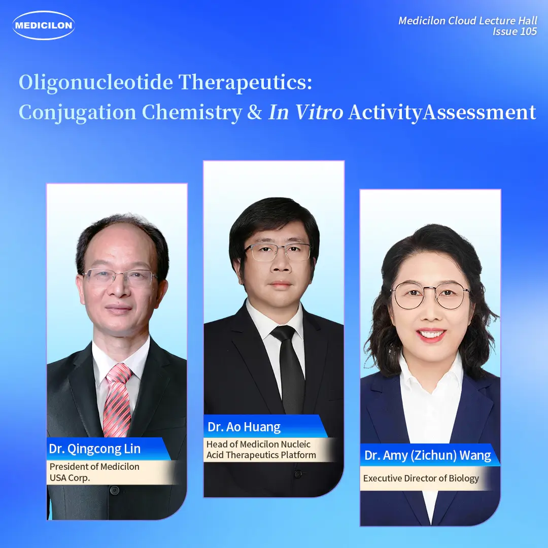
Oligonucleotide Therapeutics: Conjugation Chemistry & In Vitro Activity Assessment
- Events
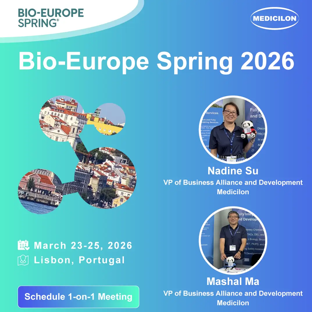
BIO-Europe Spring 2026
- Events
AACR 2026
- Events
DDC 2026
- Events
SOT 2026
Medicilon Academy
- Medicilon Academy
【Case Study】New Approach Methods NAMs
- Medicilon Academy
【Case Study】Route, Solid-State, and Impurity Optimization (Small-Molecule API)
- Medicilon Academy
【Case Study】IND Completion in 12 Months (Small-Molecule NCE)
- Medicilon Academy
Boston Non-Tumor Models Innovation
- Medicilon Academy
【White Paper】Navigating the DMPK Gauntlet: A Strategic Analysis of the Interconnected Challenges Shaping the Future of Drug Development
- Medicilon Academy
【White Paper】Patient-Derived Xenograft Organoids: Advancements and Applications in Precision Oncology
Digital Materials
- Digital Materials
Boston Innovation Meets ADME Precision
- Digital Materials
Medicilon—Developing PDX-Derived Organoid Models for Efficacy Evaluation of Anticancer Therapies (Download)
- Digital Materials
Medicilon DMPK & Bioanalysis Services (Download)
- Digital Materials
Medicilon—High Quality Integrated Drug R&D Services (Download)
- Digital Materials